


Niko McCarty and Saloni Dattani review important things happening in the world of biotechnology and medicine.
The holy grail of cancer therapy. Roughly half of all cancers have a mutation in p53, one of the most important proteins in our body. It protects us against cancer by acting as a checkpoint: when a cell’s DNA is damaged, p53 can pause the cell to help it repair the damage, or trigger the cell to self-destruct before it becomes cancerous. Destroying mutant p53, or restoring the function of healthy p53, has therefore been something of a holy grail in cancer therapy.
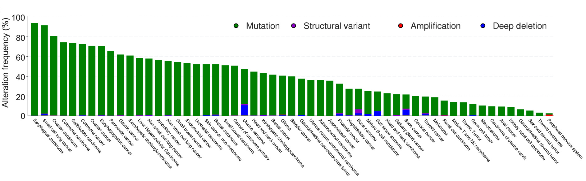
So far, though, it has been incredibly hard to develop drugs against mutant p53. That is partly because p53 can carry a wide variety of possible mutations, and partly because mutant p53 proteins tend to be slippery, without an obvious pocket where a drug could bind.

A new paper by Jennifer Doudna’s lab, which pioneered CRISPR/Cas9 as a gene-editing technology, describes a new approach. Rather than trying to attach to and block the protein, why not kill cancer cells that carry the mutation? So they developed a system to do that, CRISPR-Cas12a2. It finds mutant RNA transcripts of p53 and switches into a destructive mode, shredding the cell’s DNA, which essentially causes the cancer cell to commit suicide.
The guides are highly specific: they target just a single-letter change in the RNA and leave other cells as they are. They are delivered as mRNA inside lipid nanoparticles, with components that steer them toward the lungs, unlike the standard lipid nanoparticles that tend to be taken up in the liver. And helpfully, the nanoparticles can carry several guides at once, so one treatment could target multiple p53 mutations or cancer-causing genes together.
The first base-edited human embryo. Eight years ago, the Chinese scientist He Jiankui became infamous for using CRISPR-Cas9 to edit embryos from IVF that became children. This was more than an ethical scandal about gene-editing. The standard CRISPR-Cas9 system he used is technically risky: it cuts through both strands of DNA while making the edit, and since the break is hard for our cells to repair, it can result in losing long stretches of DNA or even entire chromosomes. Dieter Egli, a scientist at Columbia, tried it in a research setting in 2020 and found around half the embryos suffered from what he called ‘catastrophic consequences’.
Now, Egli’s group has tried another, safer approach, in the lab: base editing, which cuts only one strand of DNA instead of both, and replaces just a single letter of DNA. For certain mutations, this kind of single letter edit is enough to make a fix. The team base-edited two genes in donated embryos: PCSK9 (which regulates cholesterol levels) and HBG (which controls fetal hemoglobin), and found the edits were both efficient and free of causing the chromosomal damage seen with Cas9. But, the edited embryos weren’t implanted as He Jiankui had done.
The method resulted in some rare off-target edits, with segments aside from those of interest being edited too. And it wasn’t fully effective, as many embryos came out as genetic mosaics, with only some cells carrying the edits. (The phenomenon of being a genetic mosaic is also seen in rare cases in humans. It’s called ‘whole-body chimerism’, from two separate embryos merging into one, early in development. It’s mostly harmless and often discovered only by accident, during DNA testing, though some people with it have noticeable features like patches of differently colored skin or eyes.)
Although the technology is still in development, the idea could correct the single-letter mutations that cause many congenital diseases. Embryos found to carry such mutations during IVF, which parents might otherwise discard, could instead be repaired. Scientists have also been working on gene-editing technologies that could be applied ‘in utero’, not just during IVF, meaning gene-editing could correct congenital mutations that cause diseases like cystic fibrosis, but before birth, as fetuses grow.
The first CRISPR medicine, three years on. A few years ago, the first CRISPR medicine was approved: Exa-cel (exagamglogene autotemcel). It’s a gene-editing therapy for sickle cell disease and beta thalassemia. Both are inherited disorders of hemoglobin (the protein that red blood cells use to carry oxygen) and in both cases, the blood ends up carrying less oxygen than usual. Until recently, the only cure was a stem cell transplant from a healthy donor, but suitable matches are hard to find and transplants carry risks.
To solve the problem, the CRISPR medicine works by switching a fetal form of hemoglobin back on. People make this fetal hemoglobin naturally before birth and then switch to the adult form, and it carries oxygen just as well. In the therapy, doctors remove a patient’s blood stem cells, switch fetal hemoglobin back on with CRISPR-Cas9, clear out the remaining stem cells with chemotherapy, and then return the edited cells to the body.
A new paper reports the results of two phase 3 trials of exa-cel in children aged 5 to 11. Most children in the study were, in effect, cured: those with beta thalassemia no longer needed blood transfusions, and those with sickle cell disease became free of the painful episodes of blocked blood vessels; the remaining children hadn’t been followed up for long enough to tell. The result has led the FDA to expand the treatment to children as young as two.
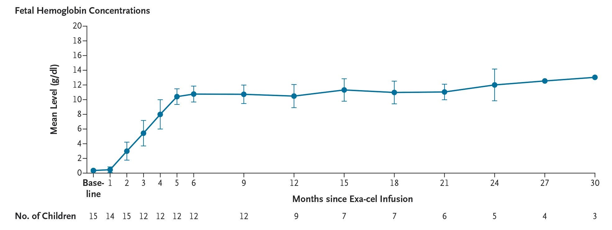
But it’s a hard therapy to go through, with side effects that can be fatal. Every child in the trials had serious side effects from busulfan, the chemotherapy used to clear the bone marrow before the edited cells go back in; two developed liver disease, and one died. The edited cells themselves weren’t harmful but the process to clear out their original cells with chemotherapy, unfortunately, was. It doesn’t have to be this way: new technologies that could skip the chemotherapy step and directly edit cells in the body are in the pipeline, though they’re currently early in development.
Lab-grown human eggs and sperm (with caveats). Two separate biotech companies – Paterna Biosciences and Conception Biosciences – have reported growing human sperm and immature eggs, respectively, in the lab.
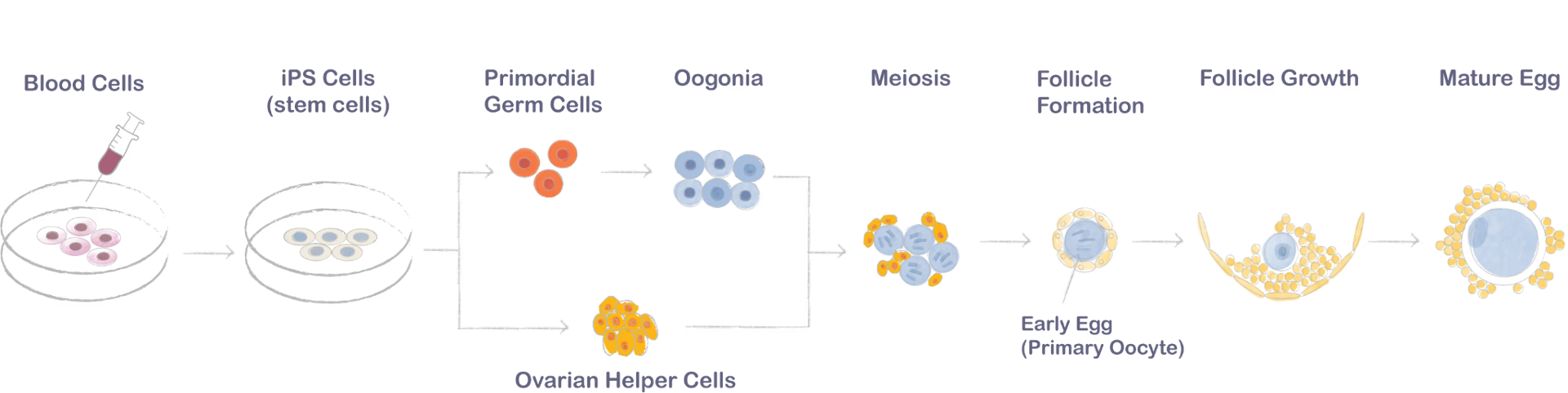
The companies are trying to achieve ‘in vitro oogenesis’, or IVG, a technology in which adult skin or blood cells are ‘reprogrammed’ back into pluripotent cells, and then coaxed to form viable eggs or sperm.1 So far, this technology has only worked in mice, with just 1–3 percent of lab grown eggs leading to viable pups. If IVG ever works for humans, though, it would mean that just about anyone who wants to have biological children could do so, including two men or two women, or women who don’t have many eggs of their own.
How about men who lack sperm? About 10 percent of men with infertility, and about 1 percent of all men, have something called ‘azoospermia’, which means they have zero detectable sperm in their ejaculate. The condition can be caused by an inability to make sperm at all, or by a blockage where sperm fail to exit the body. Paterna, one of the biotech companies working on IVG, claims to have developed a technology that would solve either problem by isolating sperm-making stem cells directly from a patient’s testicles, via a biopsy, and then coaxing them into developing mature, functional sperm in a dish. These lab-grown sperm were able to create human embryos that appear healthy, though further testing would be required to find out if the embryos had any genetic (or epigenetic) abnormalities.
The holy grail in the field, though, would be to make viable eggs or sperm all the way from blood or skin cells taken from the body, after converting the latter into pluripotent stem cells. Paterna did not do that: they are using stem cells from the testes that already have the ability to make sperm inside the body, and then recreating those sperm-making conditions outside the body.
As for the eggs, Conception Biosciences, based in San Francisco, says they’ve ‘generated the first early human egg cells (‘primary oocytes’) derived from stem cells.’ The company writes: ‘After performing a simple blood draw, we converted blood cells into stem cells, and then coaxed those stem cells into becoming miniature human ovaries that contain the early eggs.’ Note that they are only claiming to have made early eggs, which are immature, rather than mature eggs that could be used to make a human embryo.
Neither claim has been published, and experimental data is scarce. But people we’ve talked about this with in the reproductive technology field say their claims are likely real, even though it’s not clear whether either could lead to a viable pregnancy.
The race to cure multiple myeloma. Treatment for multiple myeloma, a cancer of the plasma cells in the bone marrow, has advanced a lot over the years. Much of this progress has been down to antibody treatments that direct the immune system to fight the cancer. Some, like daratumumab, tag myeloma cells so the immune system destroys them. These treatments are grueling, though: they have to be taken continuously, they carry real side effects, and in many patients the cancer relapses eventually.
Ruxandra Teslo and Amol Punjabi recently wrote about a big breakthrough from another angle: CAR-T cell therapy, where a patient’s own T cells are removed and re-engineered to hunt the cancer. The standout, Carvykti, was developed in China, and is a cure for some. About a third of patients in the phase 3 trial, with relapsed or refractory multiple myeloma, were still disease-free five years after a single infusion, with no further treatment.
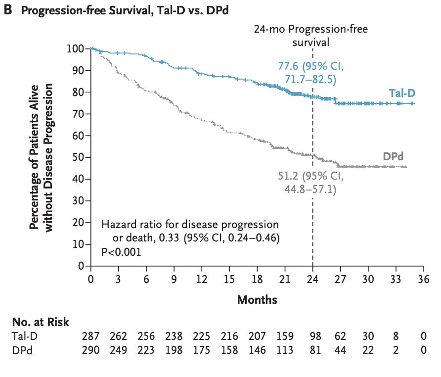
But the off-the-shelf antibodies are fighting back. Talquetamab is an example. It’s a ‘bi-specific’ antibody, which grabs myeloma cells with one arm and T cells with the other, and bridges them, helping the patient’s own T cells kill the cancerous cells. It’s been approved since 2023, but only for patients who have already been through at least four prior treatments. Now a new phase 3 trial suggests it can be given much earlier, after a first relapse.
How well does it work there? Very well! Added to daratumumab, it cut the risk of the cancer progressing by two-thirds against a standard regimen (with roughly 78 percent vs 52 percent of patients were progression-free at two years) and halved the risk of death within two years.
Electron microscopes with laser beams 100 million times brighter than the surface of the sun. Electron microscopes can help visualize objects far too small to be seen through regular light microscopes, including individual proteins. They work like this: a ‘gun’ fires electrons down a column, and magnetic lenses focus them onto a thin sample. The electrons hit the sample, scatter off, and collide with a detector, which then generates an image.
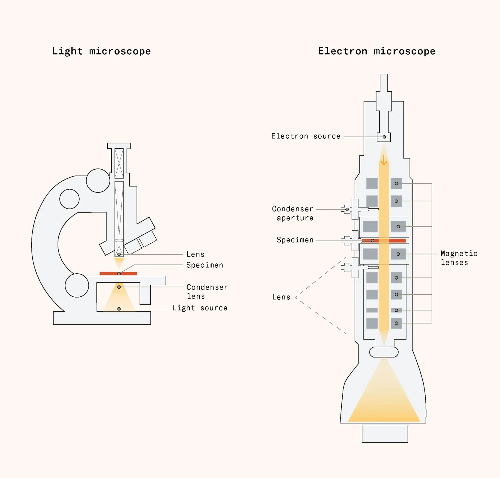
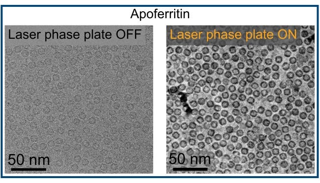
This approach has a few problems, though. For one, it only works under vacuum, so researchers must kill cells before imaging them. Even the remaining material gets ripped up and slowly destroyed by the electron beams over time. And transparent things, including cells, barely scatter the electrons passing through, so most images look blurry.
When electrons pass through a specimen, they split into two populations: most pass straight through, but a small fraction scatter, shifting their wavelength by one-quarter. At the detector, these two populations of electrons overlap and interfere, and the sum of the ‘heights’ of their waves determine the final brightness of the image. Since waves that are a quarter-wavelength apart add up at nearly right angles, the electrons scattered by the specimen have almost the same brightness as the background, which means minimal contrast.
Now, researchers at UC Berkeley have taken a huge step toward solving this problem, by using an intense laser beam to shift the phase of the electrons themselves.
First, they built a laser beam 100 million times brighter than the surface of the sun. They generated this beam by trapping a laser between highly polished mirrors, which bounce it back and forth 10,000 times to build up intensity. Then, the researchers positioned the laser so that it only interacted with the straight-through electrons, rather than the electrons that collide with the specimen, so both populations would be shifted by the same quarter wavelength. When the populations then recombine, their interference produces a large brightness difference, increasing the contrast between specimen and background.
This enables huge improvements in cryo-electron microscopy. Before, researchers could image only about 10 percent of human proteins in purified form, and under 1 percent of proteins inside intact cells, mostly larger proteins like hemoglobin. The laser phase plate could push those numbers past 50 percent, while improving their resolution too. It means researchers can see which proteins bind to each other inside living cells. They are already studying mutated lysosomes, or the recycling organelles in cells, implicated in Alzheimer’s disease.
The EU will adopt streamlined approval pathways for lightly gene-edited crops. Until now, all gene-edited crops in the EU have been regulated, regardless of how minor the edits were, under the GMO pathway. This regulatory process required many experiments – like feeding the engineered plants to rodents for 90 days to see if they got sick, proving that added proteins could not be allergenic, and mapping out all possible effects the crop could have on the environment – before approval. This process took about five years of work, and cost between 10 to 16 million euros.
But this month, the European Parliament endorsed a new regulatory system where crops carrying gene edits that are modest enough to have arisen through conventional breeding can go through expedited approval pathways. Many other countries have already adopted similar regulatory pathways that distinguish between ‘gene-edited’ and ‘GMO’ plants, including Australia, Argentina, the United States, Japan, and Brazil.
Under the new guidelines, which will go into effect in two years, engineered plants that could have been made through conventional breeding techniques will have an easier path to approval. This new pathway applies to any plants carrying less than ~20 genetic changes and with no foreign, non-plant DNA. These plants won’t need to be labeled as GMO in supermarkets, either.
Many examples of ‘gene-edited’ crops will become easier to approve in Europe as a result, including non-browning mushrooms, which are typically made by knocking out a single gene encoding an enzyme that causes that browning, and are cleared US regulatory review back in 2016. Another example is wheat resistant to powdery mildew, made by knocking out a mildew-susceptibility gene.2
A synthetic cell that can eat, grow, and divide. For the first time, scientists have made a synthetic cell (called SpudCell) that’s capable of ‘feeding, growth, replication, division and selection [...] entirely using components scientists put there.’ Although SpudCell is definitely a cell, it is not alive, because it can only divide a couple times before expiring.
It is basically a bag of lipids filled with proteins and DNA. Its genome spans 90,000 basepairs of DNA across seven plasmids. Its DNA replication is simple, using a DNA polymerase called Phi29, which floats around, randomly grabs plasmids, and copies them. This happens continuously. Its protein synthesis is more complicated, involving dozens of proteins. And SpudCell cannot make its own ribosomes; researchers must supply them from outside.
Whereas normal cells consume nutrients and convert them into useful molecules, SpudCell feeds by fusing with lipid bubbles called ‘feeder liposomes’, which are filled with ribosomes, using a protein engineered to carry short strings of histidine amino acids. These strings latch onto the feeder liposomes and coax them to merge into SpudCell to bring their ‘food’ inside.
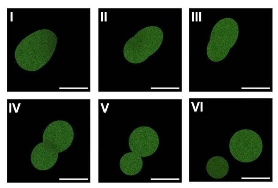
Cell division in SpudCell involves a clever trick of biophysics. It turns out that, if you stud the outside of a lipid membrane with bulky proteins, those proteins will crowd each other and repel the cell membrane, forcing the cell to bend and eventually split. And that’s how SpudCell divides.
The next step will be to engineer SpudCell so it divides dozens of times, reliably. Metabolism is probably the biggest requirement for long-term growth. As SpudCell can’t make its own ribosomes or energy, the researchers currently have to pump in fresh supplies constantly. This is fine, in principle, but SpudCell can’t recycle its broken proteins either. So when the ribosomes wear out and its proteins ‘break,’ they simply sit there and fill up space. It has no way to export its waste, so with each cell division, a larger and larger fraction of the cell’s internal volume becomes packed with junk.
Cover illustration: Escherichia coli.
.jpg)
When scientists talk about ‘reprogramming’ a cell, they mean converting it from a particular type of cell into one without an identity (termed ‘pluripotent’) so it can be transformed into almost any cell type in the body. This process is typically done by expressing four proteins in the cells, called Yamanaka factors (after their discoverer, Shinya Yamanaka), which help rewind the cells back into the pluripotent state.
This paragraph was corrected to remove the mentions of some of these products being on grocery shelves in the US – although the non-browning mushrooms been approved, they haven’t been commercialized. And non-browning, slower-spoiling potatoes, which are sold in US supermarkets today, are labeled a GMO rather than a gene-edit.
These posts are one of the highlights of my month. As someone outside the field but with a lay interest, it's so exciting to hear about all this incredible work going on, written in an a way that's accessible to non-experts. Thanks for writing them!
This is really well done! My background is mostly in physical/engineering sciences, and light on biology, but I understand these brief summaries very well. Thanks!